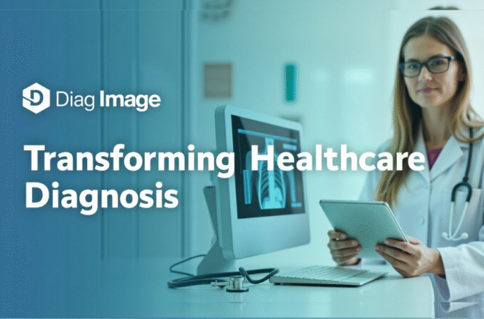
Diag imaging is the cornerstone of modern health care, which enables clinicians to look inside the human body with a clarity and precision not previously possible. Medical imaging workstations continue to play an integral role in clinical workflow, enabling clinicians to achieve accurate diagnoses and improve patient care. This is the power of the Diag image’s technology.
Based on the studies conducted across the healthcare industry, approximately 12 million Americans are subjected to diagnostic errors every year, and imaging-related diagnostic errors contribute significantly to these numbers. To meet this demand, Advanced Diag imaging technology offers radiologists and clinicians robust solutions to assist in improving the accuracy of diagnosis, while reducing interpretation errors.
Let’s explore Diag Image in more detail and try to understand how it is empowering early and accurate diagnoses.
Key Takeaways
- Diag image technology enhances diagnostic accuracy, which is crucial for patient care and reducing diagnostic errors.
- Common modalities include X-rays, CT scans, MRI, ultrasound, and PET scans, each with unique diagnostic capabilities.
- Advanced Diag image tools provide precise measurements and improve workflows for radiologists and clinicians.
- AI integration in Diag image systems increases efficiency and helps in identifying abnormalities with greater accuracy.
- Despite its advancements, Diag image technology faces challenges like costs, radiation exposure, and AI interpretation bias.
Table of Contents
- Understanding Diag Image Technology
- Common Diag Imaging Modalities
- How Diag Imaging Empowers Early and Accurate Diagnoses
- The Technology Powering Diag Image Systems
- The Role of Diag Image Centres in Healthcare Access
- Real-Life Applications of Diag Image Technology
- The Critical Importance of Diagnostic Accuracy
- Challenges and the Future of Diag Image Technology
- Conclusion
- FAQs
Understanding Diag Image Technology
Diagnostic imaging technology, often known as diagnostic imaging, refers to the procedures and equipment used to capture visual representations of the human body’s internal structures. Doctors can use these images to diagnose, monitor, and treat various medical conditions without the need for invasive procedures.
Fundamentally, the use of Diag imagery is similar to the process of looking through the window into the body, providing a clear view of soft tissues, bones, and blood vessels beyond them.
Common Diag Imaging Modalities
Let’s break down the most commonly used diagnostic imaging tests:
X-rays
These electromagnetic rays provide images of bones and other tissues in the body. X-rays help detect bone fractures, infections, and tumours.
CT Scans (Computed Tomography)
This method involves combining several X-ray images taken at varying angles to produce cross-sectional images of the body. As a result, CT scans provide detailed images of bones, blood vessels, and soft tissues. Consequently, they enable the diagnosis of cancer, cardiovascular disease, and traumatic injuries.
Magnetic Resonance Imaging (MRI)
MRIs incorporate radio frequency signals and high-power magnetic fields to create detailed images of organs and tissues. MRI is beneficial for imaging soft tissues, joints, the brain, and the spinal cord.
Ultrasound
Ultrasound involves the application of high-frequency sound waves to create images of the internal body. In obstetrics, it is used to check on the development of the fetus and examine how parts of the body are growing, such as the heart, liver, and kidneys.
PET Scans (Positron Emission Tomography)
Injects a small amount of radioactive material into the body to scan areas with a high likelihood of chemical activity. PET scans are used in oncology to detect cancer and monitor the progress of treatment.
How Diag Imaging Empowers Early and Accurate Diagnoses
Suppose we have a patient who has been experiencing continuous headaches. In addition to symptoms, a doctor may prescribe an MRI scan. The resulting Diag image may show a brain lesion that might indicate a problem such as a tumour or an aneurysm early (before it becomes life-threatening).
Precision Meets Speed
Time matters in the medical field (healthcare). Taking the example of stroke care, each minute that passes without treatment causes the brain cells to die. The Diag image technology is capable of detecting a brain bleed or clot within a few minutes; hence, treatment can be provided as soon as possible.
Minimally Invasive but Deeply Insightful
Exploratory surgery was once the only means of determining what was wrong in the body before the advent of advanced imaging. In the modern world, medical imaging provides the same level of clarity without a single cut. That means less risk, less recovery, and more peace of mind for the patient.

Advanced Visualisation and Measurement Tools
Diag image supports dedicated measuring instruments and modality-specific features for various types of images. Moreover, goniometry measurements, pan and zoom, video inversion, and high-quality image processing all help increase diagnostic confidence across a wide range of clinical applications.
| Tool Category | Key Features | Clinical Applications |
|---|---|---|
| Measurement Tools | Goniometry, distance, and area calculations | Orthopaedic assessments, cardiac measurements |
| Image Processing | Pan, zoom, video inversion, contrast adjustment | General radiology, emergency imaging |
| Display Protocols | Automated hanging protocols, layout optimization | Automated hanging protocols, layout optimisation |
| Pathological Indexing | Image cataloguing, educational database | Teaching, research, and case documentation |
These tools prove particularly valuable for complex cases requiring detailed measurements or comparative analysis. Radiologists can perform precise calculations and annotations directly within the diagnostic interface, streamlining the reporting process.
The Technology Powering Diag Image Systems
Although it might appear to be the simplest thing on the outside, a machine takes an image and displays it on a screen. The science behind Diag image technology is highly advanced.
Digital Imaging and Artificial Intelligence
Modern Diag imaging systems are digital, meaning results can be stored, enhanced, and shared across multiple healthcare platforms. Artificial intelligence (AI) is being gradually integrated into diagnostic systems, enabling radiologists to identify abnormalities, such as lung nodules or breast cancers, with greater accuracy and efficiency than ever before.
A 2020 study published in The Lancet Digital Health found that AI systems matched or outperformed radiologists in interpreting diagnostic images for breast cancer screening.
3D Imaging and Reconstruction
New developments enable 3D imaging, allowing clinicians to rotate, zoom in, and even simulate surgery planning using actual patient anatomy. This is especially useful for orthopaedic, cardiovascular, and oncological operations.
The Role of Diag Image Centres in Healthcare Access
In the United States, centres for diagnostic imaging have evolved into hubs of innovation and accessibility. These centres focus on offering a variety of diagnostic imaging tests, frequently with more specialised care and shorter wait times than hospitals..
What to Expect at a Centre for Diag Imaging
If you’ve ever visited a Diag imaging centre, here’s a quick overview:
- You will check in and respond to health-related questions.
- The technician/radiologist will explain the procedure.
- Depending on the exam, you may lie still, hold your breath, or move as directed.
- Results are generally available to your doctor within hours.
Many clinics now provide online portals where patients can quickly examine their diagnostic images and results, indicating a substantial improvement in transparency and patient empowerment.
Real-Life Applications of Diag Image Technology
Here are a few real-world examples of how Diag imaging can change people’s lives.
- Cancer Screening and Monitoring: The early stages of breast cancer are detected through mammograms and MRI (which is an X-ray). PET-CT scans help in checking the growth or shrinkage of the tumours during treatment.
- Cardiology: CT angiography also allows the identification of those coronary arteries that are blocked without any catheterisation. An echocardiogram (ultrasound) is used to assess the heart’s response to physical stress.
- Orthopaedic Injuries: MRIs and CT scans provide high-resolution images of ruptured ligaments, spinal cord difficulties, and cartilage damage, and are typically used before surgical planning.
- Neurology: MRI is the most effective way of detecting multiple sclerosis, strokes, and brain tumours. The fMRI even maps the brain functioning in a clear way, which can be used in surgery.
The Critical Importance of Diagnostic Accuracy
The financial implications of correct diagnosis are felt at various levels of medical care. Diagnostic mistakes also have high costs: malpractice costs, extra tests, and longer hospital stays. To help address these challenges, advanced Diag imaging technology delivers superior visualisation and decision support.
| Medical Specialty | Imaging Requirements | Med Diag Benefits |
|---|---|---|
| Emergency Medicine | Rapid interpretation, multi-modality access | Auto-fetching, streamlined workflows |
| Orthopedics | Precise measurements, comparative analysis | Goniometry tools, stitching capabilities |
| Oncology | Specialized measurements, protocol standardisation | Previous exam integration, advanced processing |
| Cardiology | Specialised measurements, protocol standardisation | Dedicated tools, display protocols |
It is known that computer-based diagnostic systems may decrease interpretation errors by as much as 30% for specific imaging tasks. Such systems identify and quantify potential abnormalities and provide quantitative analysis tools to assist but not replace radiologist expertise.
Challenges and the Future of Diag Image Technology
While Diag image technology is revolutionary, it isn’t without challenges:
- Cost and Insurance Coverage: Special tests like MRI or PET can be expensive. Insurance may not cover them unless they have apparent symptoms.
- Radiation exposure: CT scans and X-rays produce ionising radiation. Lower doses are safe, but recurrent exposure should be avoided unless medically necessary.
- AI Interpretation Bias: AI models rely on training data for correctness, and efforts are underway to ensure inclusivity and accuracy across different communities.
Conclusion
We may not realise it, but Diag image technology is at the root of practically every essential health technology. Diagnostic imaging and functional evaluation, as well as pain indexes, are effective mechanisms in the care of an injury. All the methods are different in the extent of detail, targeted tissue, time, cost, and risk. Your medical practitioner will consider all aspects, such as the type of medical condition you have, your past health history, and other predisposing factors, to determine whether imaging is necessary. This will help you adhere to a specific treatment plan and get well so that you can be on your feet.
FAQs
Diag Imaging solutions are designed to capture, process, and interpret medical images, so that doctors can guide patients to faster and more accurate diagnoses. It has also been tested on the X-rays, CT, MRI, and Ultrasound modalities.
Yes. Diag Imaging uses advanced algorithms and efficient workflows to help support image processing time, encourage diagnostic confidence, and overall productivity in your practice.
Absolutely. Diag Imaging combines state-of-the-art AI technology to help discover any irregularities, providing valuable information that can facilitate quicker and more accurate diagnoses.
Yes. Diag Imaging interoperates with all PACS systems to guarantee the continuous flow of diagnostic data within your hospital.
Diag Imaging has comprehensive curricula that meet clinicians’ and technicians’ needs, to guarantee all users can take full advantage of the system.


